Achilles tendon pain is a huge problem for runners. Runners have an estimated 10 fold increase risk of developing Achilles tendon pain (Achilles Tendinopathy) compared to non-runners.
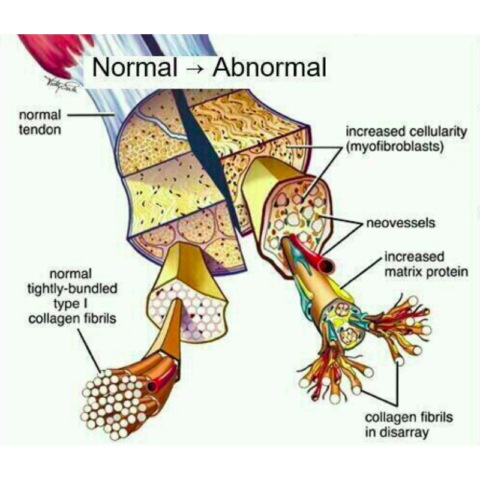
It is common for runners with Achilles tendon pain to get an ultrasound scan to see what the structure of the tendon looks like. The problem is that we don’t know what is ‘normal’ for a runner for this ultrasound scan. Changes such as collagen disorganization and reduced echogenicity mean that the tendon is showing signs of pathology. However, some of these changes may be expected in runners given how hard we work the Achilles tendon. Is it normal to see these changes in pain-free runners?
Some researchers from the University of Pennsylvania have attempted to answer that question…
Study
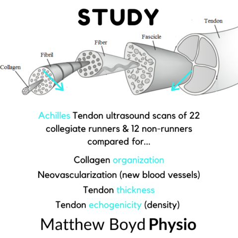
- 22 Collegiate Runners + 12 controls with no Achilles tendon pain
- Ultrasound images taken
- Collagen organization, neovascularization (new blood vessels) and tendon thickness and echogenicity (density) were evaluated
Findings
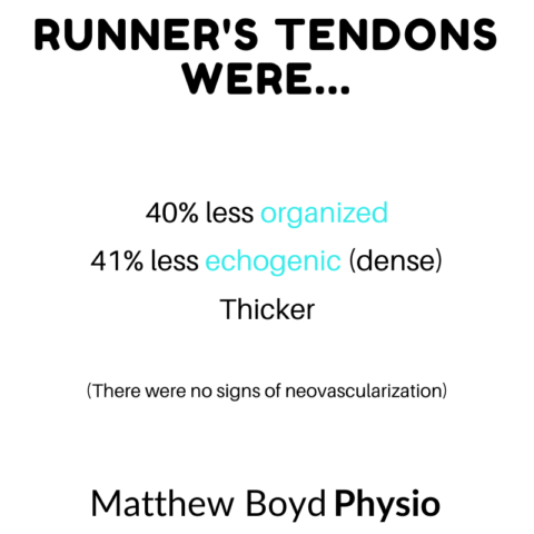
- Runners tendons:
- 40% less organized
- 41% less echogenic (dense)
- Thicker
- No signs of neovascularization
Clinicians
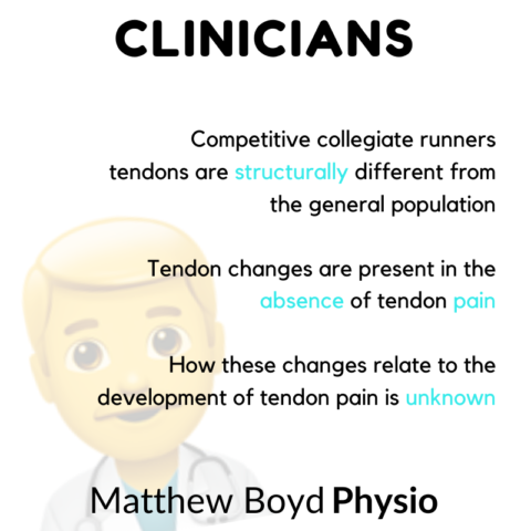
- Competitive collegiate runners tendons are structurally different from the general population
- Tendon changes are present in the absence of tendon pain
- How these changes relate to the development of tendon pain is unknown
Runners
- Your Achilles tendons probably already look different on an ultrasound scan to a non-runner
- This doesn’t necessarily mean there is a problem
Full Text
